
A technique which measures the variation in bone density within spinal bones may improve the ability to identify people at special risk of breaking their backs, Curtin University physiotherapist Andrew Briggs has found.
The method-developed by Briggs and colleagues from the Department of Medicine at the University of Melbourne and the Institute for Medical and Veterinary Sciences in South Australia-involves modifying the analysis of routine bone density x-ray (DXA) scans for bone thinning or osteoporosis to provide an assessment of bone density distribution in the back.
“We’ve shown this modification is reliable and easy to use,” Briggs say. “Now it’s a matter of demonstrating its value in a large clinical study. That would take two or three years.”
In 2001, nearly two million Australians were suffering from osteoporosis, according to Access Economics, resulting in one fracture every five to six minutes at a staggering cost of $1.9 billion, or $389 for every Australian. By 2021 Access estimates the number of sufferers will rise to three million.
Osteoporosis affects about 30% of women and 8% of men over 50. It causes a decrease in bone strength, so fractures are a common outcome. Spinal fractures account for almost half of all osteoporosis-related breaks and result in pain, mobility problems, breathing problems, postural changes, diminished strength, reduced balance and emotional disturbances.
Worse, once an initial break is sustained, the risk of further back fractures increases by between four- and seven-fold. This is known as a fracture cascade and is devastating for the patient. “While we can easily measure bone mineral density, it only provide us with an estimate of bone strength,” Andrew says. “We cannot use it to predict with confidence who is at risk of sustaining a broken back or a fracture cascade.”
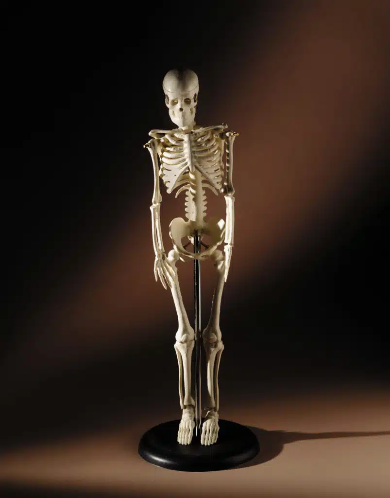
Bone mineral density is routinely measured using the DXA scanner. It’s cheap, efficient and already subsidised by Medicare for certain patient groups. But at present, DXA scans only provide a gross or ‘average’ measure of bone mineral density in the spinal bones. Such measures don’t describe how the bone mineral density is distributed within spinal bones, and this may be the key to identifying patients at risk of spine fracture.
The researchers are investigating ways to improve the routine DXA scans so the pattern of bone mineral density can be measured easily. They are now testing their techniques on donor spines before rolling out the method in a large clinical study. If successful, the technique may have major implications for clinical practice.
Andrew Briggs is one of 16 early-career scientists chosen for Fresh Science, a national program sponsored by the Federal and Victorian governments. He is presenting his research to the public for the first time at the Melbourne Museum.
Background
Dr Andrew Briggs
Australian National Health and Medical Research Council (NHMRC) Postdoctoral Research Fellow, School of Physiotherapy, Curtin University of Technology, Perth WA;
Physiotherapist, private practice, Perth, WA.
Broken backs – can we improve assessment for spinal osteoporosis?
What is the problem?
According to an Access Economics report in 2001 (Access Economics Pty Ltd, Canberra), nearly 2 million Australians had osteoporosis in 2001 and this was expected to rise to 3 million by 2021 with a fracture occurring every 3½ minutes. Osteoporosis consumed a staggering $AUD 1.9 billion in health care costs, representing $389/pa for every Australian in 2001.
The prevalence of musculoskeletal conditions increases dramatically with age, so with an aging population, the burden of these conditions is likely to become more significant both for the individual and for the Australian community. In recognition of this burden, musculoskeletal diseases, such as osteoporosis, are recognised as an Australian National Health Priority Area, while the World Health Organisation and United Nations declared the 21st century as the Decade of Bone and Joint Diseases.
Osteoporosis, or ‘thinning of the bones’, is one of the most common age-related musculoskeletal conditions, particularly in women. The condition affects about 30% of women and 8% of men aged over 50. Osteoporosis causes a decrease in bone strength, so fractures are the most significant outcome of the condition. Spine fractures are considered the hallmark of osteoporosis, accounting for almost half of all osteoporosis-related fractures. Spine fractures have several consequences including pain, mobility problems, breathing problems, postural changes, diminished strength, reduced balance and emotional disturbances. The direct cost (a quarter of the total cost) of these fractures in Australia was estimated at $ AUD 227 million/pa several years ago, so the figures today are likely to be greater. A major problem is that once an initial spine fracture is sustained, the risk of further fractures increases 4-7 fold. This problem is referred to as a ‘fracture cascade’ and is devastating for the patient.
Although we can measure bone mineral density easily, which gives a reasonable idea of bone strength, we cannot predict with confidence who is at risk of sustaining a spine fracture and the fracture cascade.
Working towards solving the problem
Bone mineral density is normally measured with a device called a DXA scanner. It’s cheap, efficient and subsidized by Medicare under certain requirements. However, routine DXA scans only make a gross or ‘average’ measure of bone mineral density in the spinal bones. However, we know that bone mineral density is not distributed evenly in the spine, so a gross or average measure doesn’t really describe how the bone mineral density is distributed in a patient. The distribution of bone mineral density may be the key to identifying patients at risk of spine fracture.
Andrew and his research colleagues (University of Melbourne Department of Medicine, Royal Melbourne Hospital, Victoria; and the Institute for Medical and Veterinary Sciences, South Australia) are investigating ways to improve routine DXA scans so that the pattern or distribution of bone mineral density can be measured easily in the clinic. The research team has proven that DXA can reliably identify differences in bone density distribution within spinal bones. They have also collected preliminary data which suggests that the technique can better identify patients who have sustained spinal fractures than routine DXA methods. The team is currently testing the technique on donor spines before rolling out the method in a large clinical study.
If successful, the technique may have major implications for clinical practice in the filed of osteoporosis.
 Fresh Science is on hold for 2022. We will be back in 2023.