
New techniques reveal how to copy nature and make bacteria-killing surgical instruments
Monday 12 February 2018
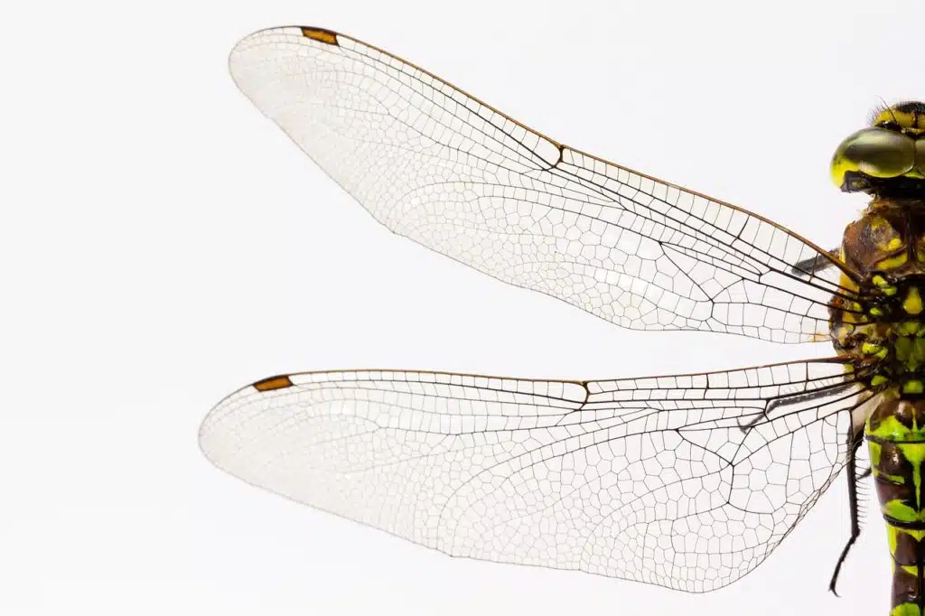
Scientists have revealed the intricate detail of how dragonfly wings kill bacteria, thanks to new methods for using very powerful microscopes to see nature’s smallest structures in three dimensions.
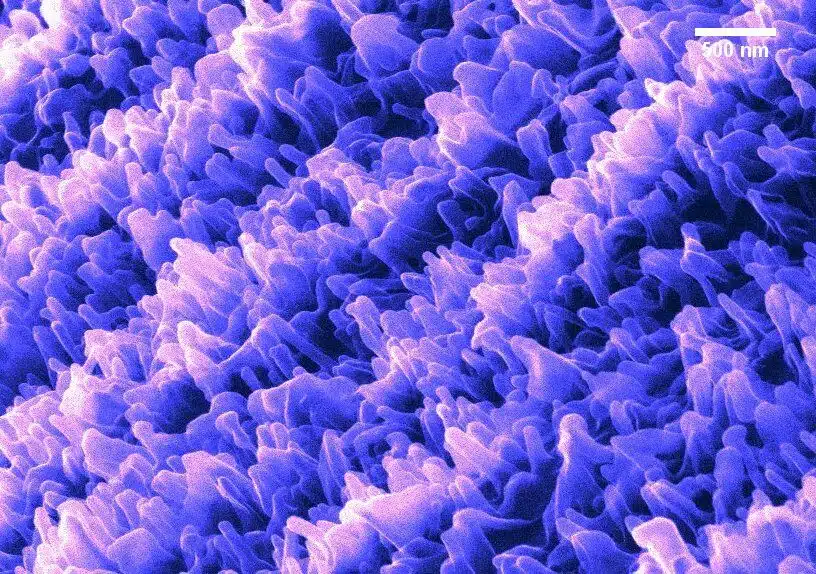
Queensland University of Technology (QUT) researchers found that dragonfly wings trap bacteria in the more than 10 billion very tiny ‘fingers’ (nanostructures) lining their surface.
“While trying to escape, the bacteria literally tear themselves apart,” says QUT researcher, Dr Annalena Wolff. “The ‘fingers on the wings are so small that even one million of them, put side by side, would be only just as large as a wasp.”
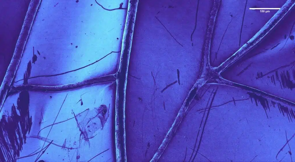
Dr Wolff says they developed new techniques for using very powerful ion and electron microscopes to look closely at tiny delicate natural structures, which may only be a few hundred atoms wide.
In the past, these microscopes easily burnt biological material, but Dr Wolff developed new techniques to overcome this problem.
“It is incredibly exciting to jump this hurdle,” says Dr Wolff. “It means we can see things that were impossible to visualise before.”
For researchers tackling deadly antibiotic-resistant bacteria, the dragonfly wing revelation offers potential new methods of dealing with the problem.
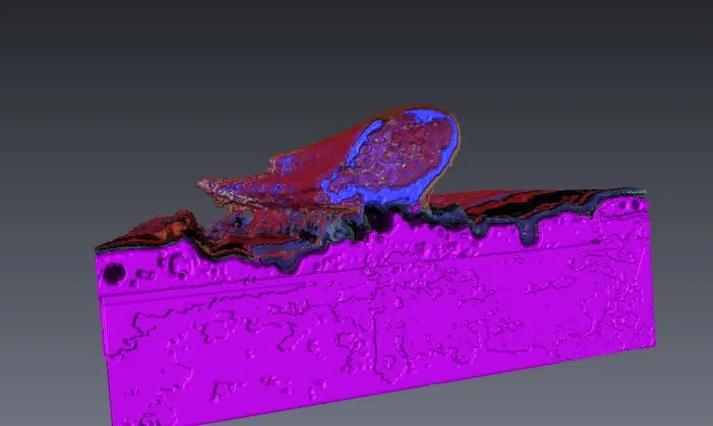
QUT researcher Dr Chaturanga Bandara says: “We have been working to unlock the secret of how dragonfly wing nanostructures destroy bacteria, so that they could be actively mimicked to surfaces of next generation smart materials, especially for use in medical applications.
“Seeing the natural nanostructure and its bacterial interaction in 3D is providing us with new clues on such new surface designs”.
Dr Wolff thinks the new images of the dragonfly wings’ nanostructures will help researchers to design things like new bacteria-killing surgery tools and hospital equipment.
“Nature has a lot of fantastic tricks up its sleeve, like the antibiotic features of dragonfly wings,” she says, “Now we can see them, and understand why they work.
“The huge microscopes we work with are two metres tall and two metres wide and can take images of almost anything.
“We can use the same microscopes to build things. For example, rebuild the dragonfly wing ‘fingers’ using different materials or build robots so small that you could fit 64 billion of them in a single raindrop. You are only limited by your imagination.”
Dr Wolff won the 2017 Fresh Science Judge’s Award (Southeast Queensland) for her work – the competition is national, and helps early-career researchers find and share their stories of discovery.
Fresh Science takes up-and-coming researchers with no media experience and turns them into spokespeople for science, giving them a taste of life in the limelight, with a day of media training and a public event in their home state.
Fresh Science Southeast Queensland is presented by Science in Public, Econnect Communication, the Queensland Government, Queensland University of Technology, the University of Queensland, Griffith University, and the University of the Sunshine Coast.
For interview
Dr Annalena Wolff, FIB/SEM Senior Laboratory, Queensland University of Technology, 0432 504 060, annalena.wolff@qut.edu.au
Dr Chaturanga Bandara, 0415052311, chaturangab@yahoo.com
Media contact
Jenni Metcalfe, Econnect Communication (for Fresh Science), jenni@econnect.com.au, phone 0408 551 866
Paper to reference
Bactericidal Effects of Natural Nanotopography of Dragonfly Wing on Escherichia coli, C.D. Bandara, S. Singh, I.O. Afara, A. Wolff, T. Tesfamichael, K. Ostrikov, A. Oloyede, ACS Applied Materials and Interfaces 2017 9 (8) 6746-6760
More about the work
Dr Wolff’s previous ScopeTV story: https://myplay.video/video/2H6WiXAncoA/scope-tv-nanobots
Videos and photos
Photos available at: www.dropbox.com/sh/ajnw42qvj1nzann/AAAsru9ZH5qRlrr_DrqtEYb-a?dl=0
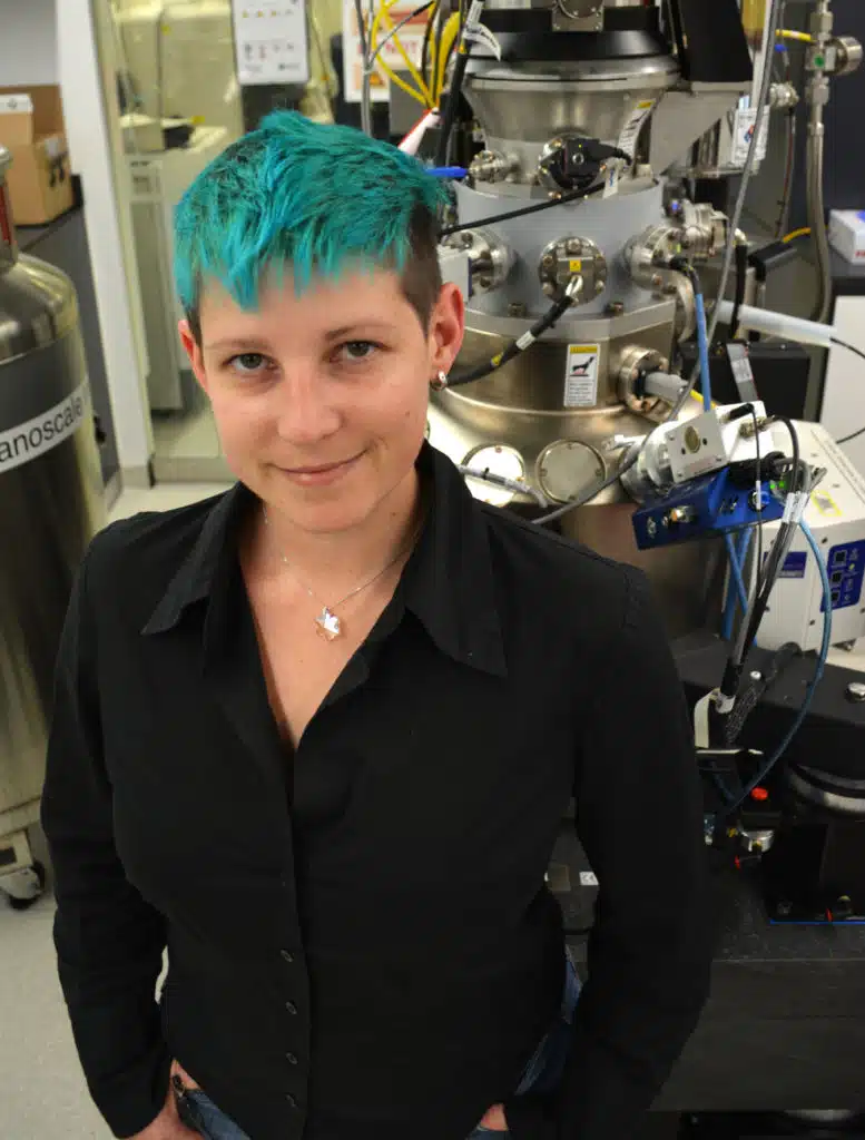
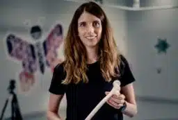
Annalena Wolff

 Fresh Science is on hold for 2022. We will be back in 2023.