
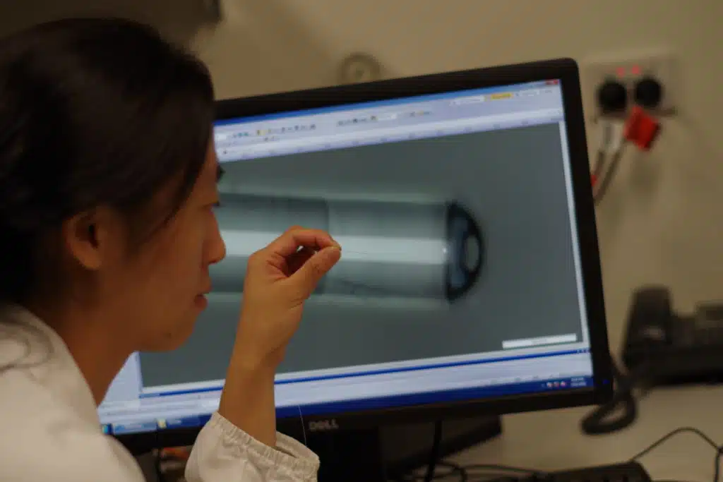
Adelaide researchers have invented the world’s smallest fibre-optic probe that can simultaneously see and measure temperature deep inside the human body.
“With an outer diameter of only 130 microns the probe is as thin as a single strand of human hair,” says Dr Jiawen Li from the Institute of Photonics and Advanced Sensing at the University of Adelaide, who was part of the research team. The research has been published in the journal Optics Letters.
“This means it can be delivered deep inside the body in a minimally-invasive way. It also allows us to see and record physiological data in real-time that we weren’t able to access before.”
Jiawen says the probe could be used to help study drug-induced hyperthermia.
“Abusing some drugs can make certain brain regions overheat and then become damaged,” she says. “Using the probe’s imaging function during experiments, medical researchers would be able to see deep inside the brain of a living organism and guide the placement of the probe to the right brain region.”
“Then, they can use the probe’s built-in thermometer to monitor any changes to the local temperature of that region.”
This will allow researchers to better understand how hyperthermia develops.
The team is also looking at ways to use the probe in other parts of the body, to make temperature-based cancer therapies safer and more effective. This is because the probe will be able to track local temperature changes in the body more accurately than existing clinical probes.
While the first generation of the probe can take both images and measure temperature, Jiawen hopes future generations will take other measurements as well – such as pH levels, oxygen saturation and accumulations of fat in the body.
Measuring fat holds exciting potential for improving our understanding of atherosclerosis, which causes heart attacks.
“The ability to study diseases or disorders where they are developing in the human body has huge implications in our ongoing search for knowledge about how diseases progress and how we can better treat them,” says Jiawen.
Contact: Jiawen Li, University of Adelaide, jiawen.li01@adelaide.edu.au
Banner image: Jiawen holding the probe. (Credit: University of Adelaide)
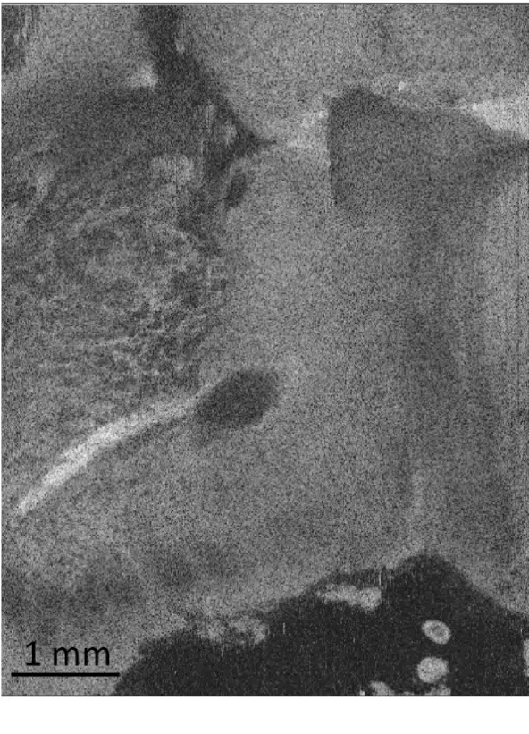
An image of a rat brain taken by the probe clearly showing different types of brain tissue. (Credit: Jiawen Li)

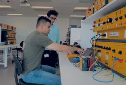
Jiawen Li presenting her research at Fresh Science SA 2017.
Jiawen Li


 Fresh Science is on hold for 2022. We will be back in 2023.