
The cells in our bodies talk with each other. Tapping into their communication could give an early warning of heart disease according to new research by Katharina Gaus, a researcher from the University of New South Wales.
One of the toughest missions in combating heart disease is an early diagnosis to allow effective treatment or surgery.
A better understanding of how cells communicate and how cholesterol levels influence this communication may prove to be one way of overcoming this problem, according to University of NSW researcher, Dr Katharina Gaus.
Next year Dr Gaus will be screening cells from heart disease patients and comparing the lipid structures from these cells with those taken from people currently unaffected by heart disease. She believes this could lead to a diagnostic test that uses cells rather than blood to determine heart disease risk in patients.
“Too much cholesterol can undermine the communication centres on the cells and threaten the way cells communicate,” she says. “Such damage happens long before our arteries are clogged up, so monitoring the state of cell communication could give us an early clue into the health of our cells and our hearts.”
The problem with monitoring a cell’s communication is that so many messages are coming in and going out that no existing computer program can simulate them.
Instead Dr Gaus has studied the process and structures involved in cell communication.
“Rather than listening to individual messages (and then having to interpret whether it is chit-chat or an emergency signal), we can monitor the communication centres to see which ones are running hot,” she says.
Cellular communication centres are sitting on the cell surface and have a characteristic lipid structure (also known as lipid rafts).
“Our new microscopy technique looks at these lipid rafts to identify and monitor any changes to their structure,” Dr Gaus said. “For example, white blood cells eat up excess cholesterol and it’s likely that high levels of cholesterol change the structure and function of lipid rafts in these cells.”
Monitoring cells in action is important because these centres are very flexible and can be rapidly assembled when needed. Because of their transient nature there has been doubt whether these lipid structures really exist but by visualising them Dr Gaus’ team has provided solid evidence for them.
Dr Gaus is presenting her research to the public for the first time thanks to Fresh Science, a national program to bring public attention to the remarkable unsung achievements of young Australian scientists. She will be speaking to the public and school students about her work on Tuesday 19 and Wednesday 20 August at the Melbourne Museum.
Katharina GausARC Post-Doctoral Discovery Research Fellow, Macrophage Biology GroupSeeing is believing: Cellular communication centres in action
Visualizing the role of lipid domains for cell signalling and activation
Every living cell in our body senses and communicates with their surrounding environment world using its surface ‘skin’ or plasma membrane. Many of these sensory events occur in specialised patches on the plasma membrane, called ‘lipid rafts’. We have developed for the first time a microscopic method to allow us to look directly at lipid rafts floating on the cell surface.
Project description
Cell membranes are the walls that separate the contents of every cell from the outside world. They are made of an oily skin of fat molecules (lipids) into which a number of large proteins are stitched. We have known for some time that many of these membrane proteins act as antennae to receive or transmit signals to the outside world. On the other hand, until recently the lipids were considered simply as inert, water-insoluble bricks in the wall, with little or no active role in the life of cells. However, this has now changed…
Like ourselves, each cell in our body needs to communicate with its environment and neighbours. This all takes place across the cell surface membrane. We can think of cell surface proteins as telephones (with call-forward facilities) that receive calls, take messages and redirect them to other parts of the cell. For example, when a growth factor receptor protein on the cell membrane senses high levels of growth factor nearby, it transmits this signal, through a chain of other proteins inside the cell, to the nucleus, to tell the to cell divide. As for telephone calls, where callers are connected through a central telephone exchange, so the cell membrane brings many of the proteins that are talking (signalling) to each other closer together, to increase the speed and accuracy with which messages can be sent. It does this by using specialised fat patches on the cell membrane. These patches are much more rigid than the surrounding membrane and so are called ‘lipid rafts’, which float on the ‘sea’ of membrane oils. Many of the proteins that receive and transmit signals are gathered together on these lipid rafts.
While the existence of lipid rafts was only recently discovered, we already know that they are very dynamic structures, constantly changing their protein cargo to respond to the changing cell environment. We know that they are important in many reactions of cells. For example, lipid rafts are the places on cells where growth factors trigger cell division, where cells of the immune system communicate with each other and where nitric oxide (the chemical that controls blood pressure and erection) production is controlled. Lipid rafts are also concentrated on membranes white blood cells use to engulf bacteria and are taken over by some viruses to invade cells and to replicate. So clearly they play an important role in biology.
But there are lots of things still to learn. For example, although we know cholesterol is an important fat in lipid rafts, we have no idea whether people with high blood cholesterol have unusual numbers or types of lipid rafts and what impact this might have on their health. What we have done is to develop a method that allows us to look directly at lipid rafts in cell membranes. We have exploited the difference in the rigidity of lipid rafts and other parts of the membrane, using a dye that changes colour depending on fluid structure. Using a special microscope we can look directly at living cells to see the distribution of lipid rafts and to watch how this changes as the cell adapts to different conditions.
By using this new method, we will be able to get a more detailed insight into how lipid rafts control cell communications. As we learn their language we might be able to take better care of them – after all they are doing a mind-boggling job for us.
Personal details
Qualifications: Centre for Vascular Research, UNSW, Ph.D. (1999) University of Cambridge, UK; M.Phil (1996) University of Cambridge, UK Centre for Vascular Research, School of Medical Sciences, Centre for Vascular Research, School of Medical Sciences,
Address: Macrophage Biology Group Centre for Vascular Research, School of Medical Sciences, University of New South Wales Sydney 2052 NSW.
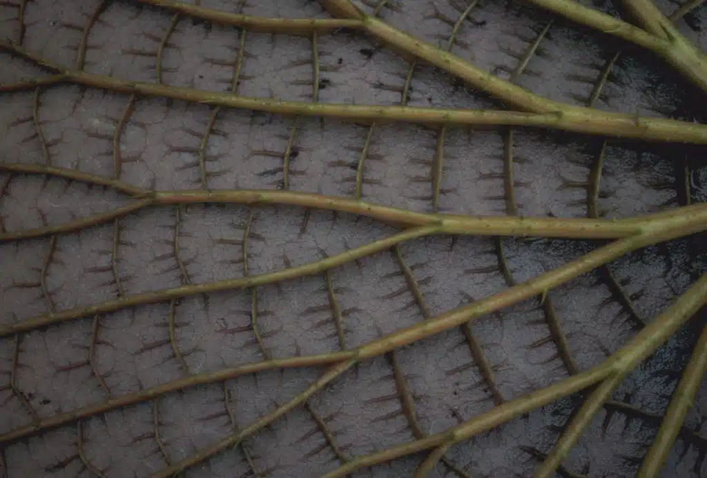
Please click the thumbnail for a high-res image
|
3-D image of a cross-section of a living macrophages (a type of white blood cells) with active communication centre in yellow/red . One can see that the communication centres (lipid rafts) are near the surface of the cell and also sometimes in the ‘spikes’ which are used to make contact with other cells and/or attach bacteria. |


 Fresh Science is on hold for 2022. We will be back in 2023.