
A new brain tumour model to provide more realistic test responses to chemo and radiotherapy is being developed by NSW scientists.
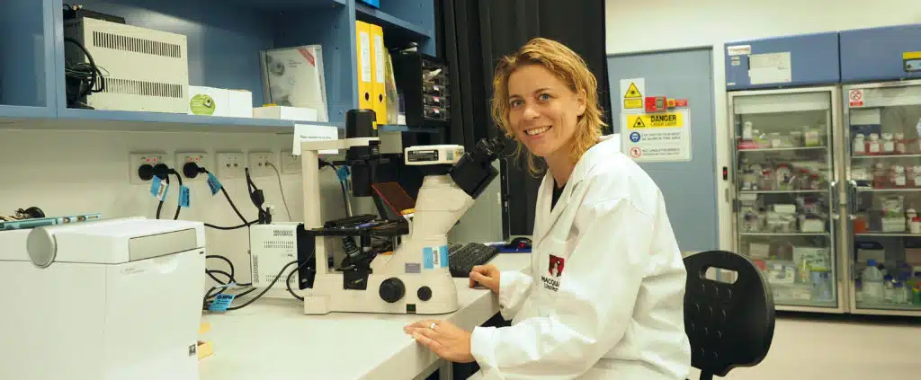
“High-grade brain cancer is very aggressive and treatment outcomes have not improved much for several decades,” says Dr Annemarie Nadort of Macquarie University, who is leading the research.
“One of the reasons for this delay is the lack of a lab-based model that accurately reflects the tumour environment in the brain.”
Current tumour models are grown in plastic Petri dishes, whereas the new models use a soft cushion of brain-like tissue as a scaffold for the tumour cells to grow on—making the testing environment more like real life.
The scaffolds are derived from animal brain materials, sourced fresh from the butcher and washed until only the structural material is left.
The team can make up to 100 mini brain tumour models from one animal brain, making it a sustainable way for high throughput drug screening.
“The effects of both chemotherapy and radiotherapy can be measured using this model, as with previous models—but it will provide a more realistic environment,” Annemarie says.
The new model will also be used to develop new tools for detecting tumour margins during neurosurgery, in a collaboration with Dr Andrew Davidson from the Macquarie University Hospital.
It’s hoped that these projects will allow for faster feedback on therapy, and more complete tumour removal, ultimately improving the quality of life for patients.
Banner image: Dr Annemarie Nadort studies mini 3D brain tumours to improve cancer therapy. Credit: Tony Crawshaw
 Fresh Science is on hold for 2022. We will be back in 2023.